The Knee Joint
Home
Anatomy
Function
Prevention
Injuries and Surgeries in Football
Stretching Exercises
Strengthening Exercises
References
Anatomy Of the Knee Joint
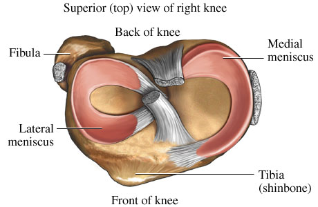
Bones
Femur
The femur is a long bone and it is found in your thigh
The distal ends of the femur form the condyles which articulate with the tibia and patella
This is a weight bearing bone
Tibia
The tibia is located on the medial aspect of your lower leg
It is a weight bearing bone
Fibula
The fibula is located on the lateral aspect of the lower leg
It is not a weight bearing bone but rather serves as an attachment site for other structures.
Patella
The patella is the largest sesamoid bone in the body.
This bone is more commonly known as the knee cap.
The patella is located on the anterior aspect of the knee joint and articulates with the femur and the tibia
It is held in place by the patellar tendon and the quadriceps tendon
Menisci
The menisci attach to the tibial plateaus within the knee joint
The meniscus is supplied blood by the medial genicular artery
The outer portion, peripheral one-third, of the meniscus has a good vascular supply but the inner one-third is avascular
Medial Meniscus
C-shaped fibrocartilage
Lateral Meniscus
O-shaped fibrocartilage
This is more smaller and mobile than the medial.
Ligaments
Anterior Cruciate Ligament (ACL)
Ligament is within the joint capsule
Comprised of 3 twisted bands
Attaches below and in front of the tibia
Attaches laterally to the inner surface of the lateral condyle
The ACL works together with the hamstring muscle group to stabilize the knee joint
Posterior Cruciate Ligament (PCL)
Ligament is within the joint capsule
Stronger than the ACL
Crosses from the back of the tibia
Attaches to the anterior portion of the lateral surface of the medial condyle
The PCL is taut throughout full range of motion (ROM)
Medial Collateral Ligament (MCL)
On the medial outer capsule of the knee joint
Moves in synchrony with the LCL
Attaches on the medial epicondyle
Inserts on the tibia below the pes anserinus
Lateral Collateral Ligament (LCL)
About the size of a pencil
Attaches to the lateral epicondyle
Inserts on the head of the fibula
Taut during knee extension
Musculature
Hamstrings
Biceps Femoris
Semitendinosus
Semimembranosus
Quadriceps
Rectus Femoris
Vastus Lateralis
Vastus Medialis
Vastus Intermedius
Sartorius
Originates at the ASIS
Inserts on the proximal medial surface of the tibia
Popliteus
Originates at the lateral condyle of the femur
Inserts on the posterior surface of the tibia
Gastrocnemius
Has two heads, lateral and medial head
insert on the posterior surface of the calcaneous, Achilles Tendon
Gracilis
Originates at the inferior ramus of the pubis
Inserts on the medial surface of the tibia
Plantaris
Originates on the lateral supracondylar ridge of the femur
Inserts on the posterior surface of the calcaneous
Bursae
Bursa are comprised of synovial tissue and thin films of fluid
Essentially are sacks of fluid and are found throughout the body
Possibly 24 bursae have been identified around the knee joint
Most common are: suprapatellar, prepatellar, and infrapatellar
Nerve and Blood Supply
Tibial nerve innervates the hamstrings and gastrocnemius
The common peroneal nerve innervates the short head of the biceps femoris, popliteal fossa and wraps around the proximal end of the fibula
Femoral nerve innervates the quadriceps and sartorius
The knees main blood supply is from the popliteal artery, stems from the femoral artery.
Blood returns back to the heart by the small saphenous vein, to the popliteal vein, to the femoral vein
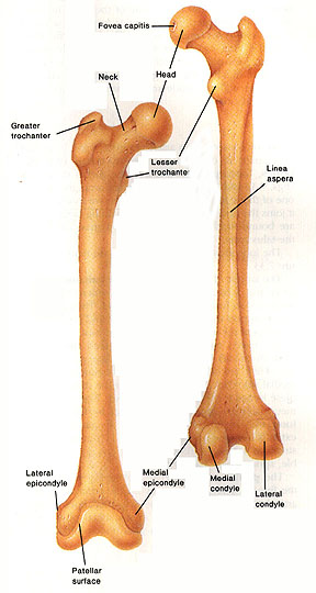
Image copied from: http://www.edoctoronline.com/medical-atlas.asp?c=4&id=22094
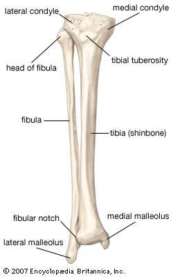
Imaged copied from: tibia: human tibia and fibula. Art. Encyclopædia Britannica Online. Web. 9 Apr. 2013.
.
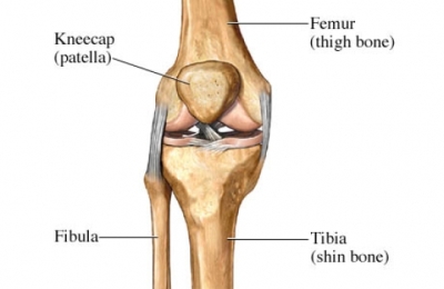
Image copied from http://www.aurorahealthcare.org/yourhealth/healthgate/images/si55550925.jpg
http://deansomerset.com/2011/01/27/the-best-exercises-you-could-ever-do-quad-activation-progressions/
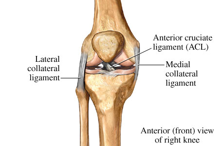
Image copied from http://www.webmd.com/pain-management/knee-pain/ligaments-of-the-knee-anterior-front-view
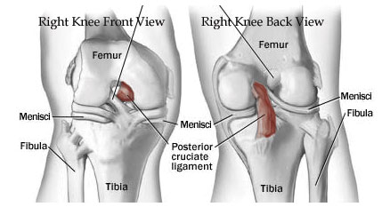
Image copied from http://www.redmondpt.com/RedmondPTPCL.html
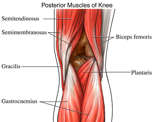
Image copied from http://iv.nucleusinc.com/generateexhibit.php?ID=4337&A=1029
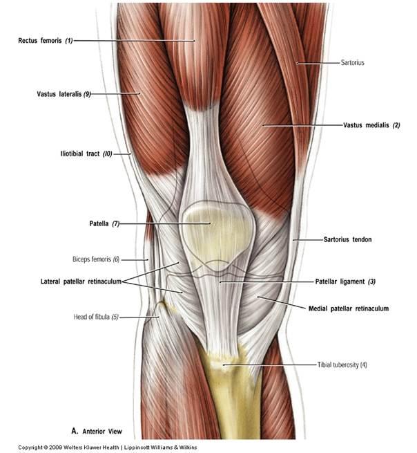
Image copied from http://classconnection.s3.amazonaws.com/694/flashcards/597694/jpg/patella1312149569783.jpg
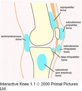
Image copied from http://solomonsseal.wordpress.com/2010/04/28/treating-bursitis-of-the-knee-with-solomons-seal/